BrainVoyager v23.0
Removing MP2Rage Background Noise
The MP2Rage (magnetization prepared 2 rapid gradient echoes) sequence is often used at 7 Tesla MRI scanners to get T1-weighted anatomical measurements with very good grey-white matter contrast. It typically results in a series of 3D volumes containig different weightings, including the UNI
, INV1
and INV2
files. The UNI volume is supposed to be used for further processing but its background voxels exhibit high intensity values which makes subsequent processing and visualization difficult. BrainVoyager offers a simple tool to denoise
the UNI volume that is based on the publication by O'Brien et al. (2014). The resulting denoised UNI volume appears with a rather clean background as one might know from conventional T1w sequences such as the MPRage. The O'Brien et al. (2014) denoising technique does not require availability of phase images but one needs to provide the INV1
and INV2
images containing the first and second inversion times.
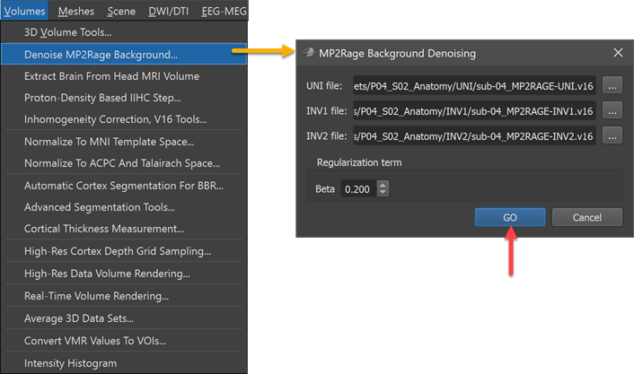
In order to remove the high-intensity background noise, the new MP2Rage Background Denoising dialog can be used. The dialog will launch after clicking the Denoise MP2Rage Background item in the Volumes menu (see screenshot above). The required VMR/V16 files can be selected using the respective Browse buttons on the right side of the UNI file INV1 file and INV2 file text fields. The screenshot above shows the dialog after the respective files from a MP2Rage scan have been selected. The calculations to denoise the background use a regularization term that can be changed but it is recommended to keep the default value of '0.2' (O'Brien et al., 2014). In order to produce the denoised VMR-V6 output file, the GO button needs to be clicked.
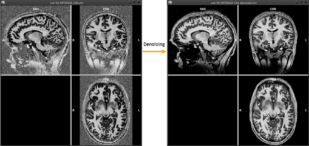
The screenshot above shows the original UNI volume on the left side and the resulting denoised output volume on the right side. The name of the produced output UNI file is based on the name of the input UNI file with the added substring _denoised
(see e.g. the VMR names in the title bars of the two volumes in the screenshot above). As one can see in the example, the output volume has been indeed cleaned
, i.e. its background voxels have low intensity values as indicated by the black color.
References
O'Brien KR, Kober T, Hagmann P, Maeder P, Marques J, et al. (2014). Robust T1-Weighted Structural Brain Imaging and Morphometry at 7T Using MP2RAGE. PLoS ONE, 9(6): e99676.
Copyright © 2023 Rainer Goebel. All rights reserved.