BrainVoyager v23.0
Retinotopic Mapping of Early Visual Areas
Background
Retinotopic maps are based on topography-preserving projections from the retina to the LGN layers and from the LGN layers to area V1. While the size of receptive fields increase from V1 to V2, V3 and V3A/V4v, these areas also show a retinotopic organization. Using fMRI and appropriate visual stimuli, early visual areas can be charted in individual human brains (Sereno et al., 1995). Retinotopic mapping experiments can be analyzed in volume (FMR and VMR) space (figure 1A) or folded cortex meshes (figure 1B). In order to reveal, however, the topological nature of these maps, retinotopic mapping data should be analyzed and visualized on flattened cortex representations (figure 1C,D).
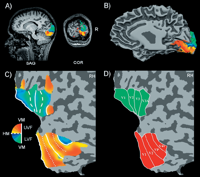
Figure 1 (reprinted from Goebel et al. 2003).
Retinotopic mapping is possible because the mapping from the retina to the primary visual cortex is topographical and because other early visual areas retain this retinal topography, but represent the visual field in either a mirrored or nonmirrored two-dimensional layout. In a topographical mapping, neighboring regions in one area (retina) project to neighboring regions in another area (primary visual cortex). Since this mapping is preserved also at the next cortical stages (V2, V3/VP, V3A, V4), neighboring positions in the visual field tend to be represented by groups of neurons that are adjacent but laterally displaced within the cortical gray matter of these areas. This transformation preserves the qualitative spatial relations of the retina but exhibits systematic distortions, i.e., there is much more cortical tissue devoted to foveal regions than peripheral regions of the visual field (cortical magnification). A successful delineation of the early visual areas with fMRI exploits the fact that successive visual areas alternate with a mirror or nonmirror representation of the visual field. Multiple horizontal and vertical meridian representations across the visual cortex, thus, indicate the transitions between early visual areas. With appropriate visual stimuli, fMRI studies are able to reveal multiple meridian representations arranged in approximately parallel bands along the flattened cortical surface (see figure above). fMRI-based retinotopic mapping has quickly become an important tool to noninvasively define the borders of early visual areas (e.g., Tootell et al., 1996, 1997; Goebel et al., 1998, 2001, 2003).
In the following it is assumed that retinotopic mapping experiments have been performed with a similar experimental procedure as described in Sereno et al. (1995), DeYoe et al. (1996) or Goebel et al. (1998). If you plan to do such experiments in the future, you may download our retinotopic mapping software from our web site (coming soon). As a minimum prerequisite, you need to map the horizontal and vertical meridians in order to be able to manually delineate boundaries on cortical flat maps. The following description assumes that you have run full eccentricity and polar angle experiments with an expanding (and/or shrinking) checkerboard ring stimulus and a rotating checkerboard wedge stimulus, respectively. These experiments are also code "phase-encoding" experiments because stimulation of the visual field is repeatedly performed and the response to a specific eccentricity or polar angle corresponds to the phase (relativetime point) within a cycle.
In the next section, the basic analysis of phase-encoded retinotopic mapping experiments will be described. The resulting boundaries between early visual areas may be highlighted by defining Patches-Of-Interest (POIs). After proper treatment (including spatial smoothing) of the obtained retinotopic maps, boundaries between early visual areas may also be determined by the computation of field sign maps (Sereno et al., 199?).
References
Goebel, R., Khorram-Sefat, D., Muckli, L., Hacker, H. and Singer, W. (1998) The constructive nature of vision: Direct evidence from fMRI studies of apparent motion and motion imagery. The European Journal of Neuroscience, 10, 1563-1573.
Goebel, R., Muckli, L., Zanella, F.E., Singer, W. & Stoerig, P. (2001). Sustained extrastriate cortical activation without visual awareness revealed by fMRI studies of hemianopic patients. Vision Research, 41, 1459-1474.
Goebel, R., Muckli, L. & Kim, D.-S. (2012). The Visual System. In: G. Paxinos & J.K. Mai (Eds.), The Human Nervous System, 3rd Edition. New York: Academic Press.
Copyright © 2023 Rainer Goebel. All rights reserved.