BrainVoyager QX v2.8
Segregation of Hemispheres
After segmentation of the white / grey matter boundary, the two hemispheres are segregated next if the Disconnect hemispheres option is turned on (default).

In this step, a cut is performed within the midsagittal slice (x = 128) separating the brain in the region of the corpus callosum and subcortical structures (90 < y < 175, 90 < z < 150). This cut is not performed along the whole midsagittal slice because this could introduce wrong results due to asymmetries of the two hemispheres. Such asymmetries are often present in the occipital lobes where one hemisphere often extends across the midsgittal plane into the region of the other hemisphere. Therefore, the initial cut through the mid-sagittal slice excludes posterior parts of the brain; a subsequent region growing process in each hemisphere is used to find the boundaries of the two hemispheres in the occipital lobe. Only if this attempt fails, the more simple method of cutting through the whole midsagittal plane is used and the user is informed about this with a message dialog. It is recommended to check the resulting hemisphere separation in this case and to manually improve the result if necessary.

During the disconnection step, the two segmented hemispheres are shown in two colors as depicted in the figure above. The segmentation is first performed on the non-dilated version of the brain and is then extended also to the dilated version (see previous step). Since the two hemispheres are segregated (highlighted with two different colors) they can be finally saved to disk separately under the names "[name]_LH_WM.vmr" and "[name]_RH_WM.vmr". If you open these files (see figure below, left hemisphere on the left, right hemisphere on the right), you will see that the colors are changed to blue (color index: 240) with a yellow boundary (color index: 235). This is because before saving the files, a process called "prepare for surface reconstruction" has been performed, which allows to use these segmentations directly for surface reconstruction of the cortical sheet.
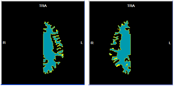
During surface reconstruction, the borders of the yellow voxels will be used to create a mesh (surface) representation of the cortical sheet.
Copyright © 2014 Rainer Goebel. All rights reserved.