BrainVoyager QX v2.8
Mask-Based Removal of Non-Cortical Structures
While Talairach normalization is not the perfect method to transform the cortex of different subjects in a common space, it does transform subcortical brain structures to quite similar spatial positions. This property of Talairach (and ACPC) space is exploited as a priori location information in the next two steps of the automatic segmentation procedure that are included if the Find and fill ventricles and Apply brain mask options are turned on (default).
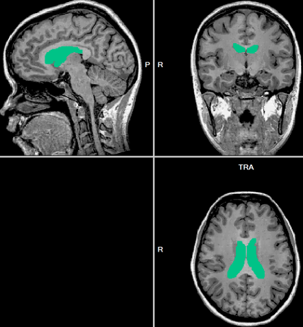
In the first of these steps, voxels with low intensity values are sampled around locations that belong with a high likelihood to ventricles. Note that ventricles have low intensity values ("black" voxels) in T1-weighted structural images. While ventricles are roughly at the same positions in different brains, their exact size and shape vary considerably across subjects. The program therefore starts a region growing process from one of the detected "black" voxels. This process labels voxels in the neighborhood having also low intensities resulting in filled ventricles up to the boundaries with high intensity values. The dark green regions in the images below depict the labeled (filled) ventricles in a brain after running this step.
In the second step, a mask is applied to the data set to roughly segment the brain from the surrounding head tissue including the cerebellum. The mask also labels subcortical structures as "white matter". Otherwise the grey/white tissue of subcortical nuclei would appear as cortical grey/white matter boundaries in the final segmentation. The coarse Talairach-based brain segmentation and labelling of subcortical structues is performed based on a VMR mask file called "BrainTalMask.vmr". This file is located in the BrainVoyager QX folder after a standard installation of the program. The snapshot below shows three orthogonal slices of this mask file. The green regions in that file are used to automatically label subcortical structures as "white matter" and the red/orange regions are used to perform an initial brain segmentation including removal of the cerebellum. These initial segmentation will be refined by subsequent steps.
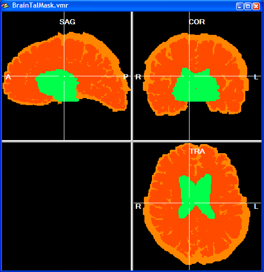
The Talairach brain mask file has been created based on the segmentation of an individual brain (dark orange color) and it was then expanded to fit many additional brains (light orange color). This resulted in a "conservative" mask file which can be applied to any new brain without removing relevant brain tissue. Since the BrainTalMask.vmr file is a standard VMR file, it is possible to change it easliy yourself, if desired. BrainVoyager QX uses the dark orange color (color index: 226) and light orange color (color index: 229) to segment the brain and the green color (color index: 245) to label subcortical structures as white matter. Applying the mask keeps the original intensity value at voxel positions at which the mask has a non-zero value and removes voxels (intensity set to 0) at positions at which the mask is black.
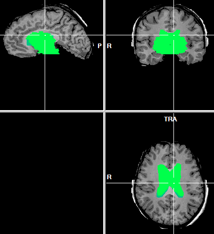
The snapshot above depicts the resulting state of the segmentation procedure after applying the mask to the brain shown above.
Copyright © 2014 Rainer Goebel. All rights reserved.